Sagittal view of nasal cavity – Embark on a journey through the sagittal view of the nasal cavity, a fascinating anatomical perspective that unveils the intricate structures and clinical implications of this essential respiratory passageway.
Delving into the nasal cavity’s sagittal view, we encounter a symphony of anatomical landmarks, including the nasal septum, turbinates, and paranasal sinuses, each playing a vital role in respiration, olfaction, and overall nasal health.
Introduction
Understanding the sagittal view of the nasal cavity is crucial for several reasons. Firstly, it allows us to visualize the intricate arrangement of anatomical structures within the nasal cavity, providing insights into their relationships and functions. Secondly, this view is essential for diagnosing and managing various nasal conditions, such as septal deviations, turbinate hypertrophy, and nasal polyps.
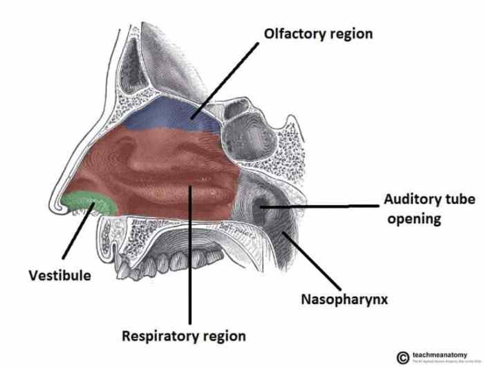
The sagittal view of the nasal cavity reveals several key anatomical structures. The nasal septum, a midline structure, divides the nasal cavity into two halves. The lateral wall of the nasal cavity is formed by the turbinates, which are scroll-like structures that increase the surface area for air filtration and warming.
The floor of the nasal cavity is formed by the hard palate, while the roof is formed by the cribriform plate of the ethmoid bone, which allows the passage of olfactory nerves.
Structures of the Nasal Cavity: Sagittal View Of Nasal Cavity

The nasal cavity is a hollow space located in the skull that allows for the passage of air during respiration. It is divided into two halves by the nasal septum, a midline partition of bone and cartilage. The roof of the nasal cavity is formed by the cribriform plate of the ethmoid bone, which contains perforations for the passage of the olfactory nerves.
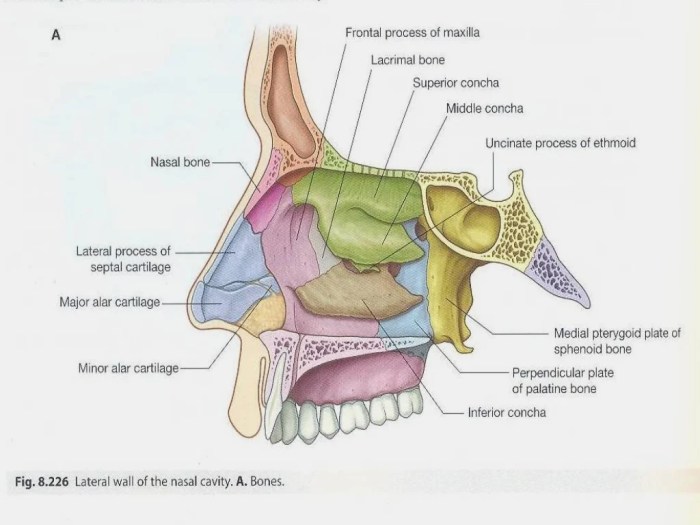
The floor is formed by the palatine processes of the maxillae and the horizontal plates of the palatine bones. The lateral walls of the nasal cavity are formed by the maxillae, the palatine bones, the inferior turbinates, and the middle and superior turbinates.
Anatomical Structures Visible in the Sagittal View
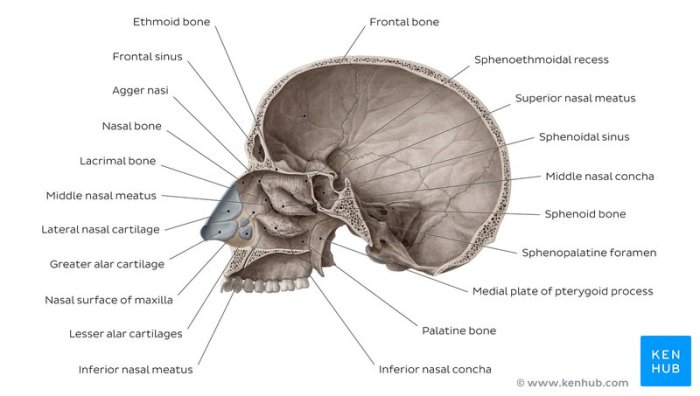
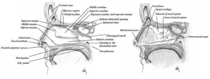
The sagittal view of the nasal cavity reveals several important anatomical structures.
The sagittal view of the nasal cavity, which offers a detailed look at its structures, can be an important reference for those seeking insights into the jp morgan superday offer rate . This view helps visualize the relationship between the nasal septum and turbinates, providing valuable information for understanding nasal anatomy and airflow dynamics.
Nasal Septum
- The nasal septum is a midline partition that divides the nasal cavity into two halves.
- It is composed of bone (the perpendicular plate of the ethmoid and the vomer) and cartilage (the septal cartilage).
- The nasal septum helps to direct airflow and prevent nasal obstruction.
Turbinates
- The turbinates are three pairs of bony projections that extend from the lateral walls of the nasal cavity.
- They are covered by a mucous membrane that helps to warm, moisten, and filter the air that passes through the nasal cavity.
- The inferior turbinate is the largest and most prominent of the turbinates.
- The middle turbinate is located above the inferior turbinate and is smaller in size.
- The superior turbinate is the smallest and most superiorly located of the turbinates.
Paranasal Sinuses
- The paranasal sinuses are air-filled cavities located within the skull that are connected to the nasal cavity.
- They help to lighten the skull, resonate the voice, and produce mucus that helps to keep the nasal cavity moist.
- The paranasal sinuses include the maxillary sinuses, the frontal sinuses, the ethmoid sinuses, and the sphenoid sinuses.
Clinical Significance

The sagittal view of the nasal cavity provides valuable insights for diagnosing and managing nasal disorders. It enables visualization of the nasal structures and their relationship to adjacent anatomical regions.
Imaging techniques such as computed tomography (CT) and magnetic resonance imaging (MRI) are commonly used to obtain sagittal views of the nasal cavity. These imaging modalities provide detailed cross-sectional images that can reveal structural abnormalities, inflammatory changes, and other pathological conditions.
Nasal Obstruction
The sagittal view is crucial for evaluating nasal obstruction, which can result from various causes, including septal deviations, turbinate hypertrophy, and nasal polyps. The sagittal view allows visualization of the nasal septum, turbinates, and nasal passages, enabling the identification of structural abnormalities that may obstruct airflow.
Sinus Infections
The sagittal view also plays a significant role in the diagnosis and management of sinus infections, such as sinusitis. It provides a clear visualization of the paranasal sinuses, allowing for the assessment of mucosal thickening, fluid accumulation, and other signs of inflammation.
This information guides appropriate treatment decisions, including antibiotic therapy or surgical intervention.
Variations and Anomalies
The sagittal view of the nasal cavity typically exhibits variations in size, shape, and the presence of anatomical structures. Understanding these variations is crucial for accurate diagnosis and surgical interventions.
Common anomalies include septal deviations, turbinate hypertrophy, and concha bullosa. These anomalies can obstruct airflow, leading to nasal congestion, sinusitis, and other respiratory issues.
Septal Deviations
- Septal deviations are misalignments of the nasal septum, the cartilage and bone that divides the nasal cavity into two halves.
- They can be congenital (present at birth) or acquired (develop later in life due to trauma or injury).
- Severe deviations can block airflow, causing difficulty breathing, nasal congestion, and facial pain.
Turbinate Hypertrophy
- Turbinate hypertrophy refers to the enlargement of the turbinates, the bony structures located on the lateral walls of the nasal cavity.
- Enlarged turbinates can obstruct airflow, leading to nasal congestion and difficulty breathing.
- Causes include allergies, chronic inflammation, and hormonal imbalances.
Concha Bullosa
- Concha bullosa is a condition in which the middle turbinate contains air-filled cavities or cysts.
- These cysts can enlarge and obstruct the nasal cavity, causing nasal congestion and sinusitis.
- It is often asymptomatic but can lead to complications such as nasal polyps and chronic sinusitis.
Surgical Approaches

The sagittal view of the nasal cavity provides valuable insights for surgeons planning surgical approaches to this region. It allows visualization of the nasal cavity’s depth, width, and the relationship between its structures.
Various surgical techniques are employed to access and operate on the nasal cavity through the sagittal approach. These techniques include:
Endoscopic Sinus Surgery (ESS)
- Minimally invasive procedure using a nasal endoscope and specialized instruments.
- Allows surgeons to access and visualize the nasal cavity and paranasal sinuses without external incisions.
- Commonly used to treat chronic sinusitis, nasal polyps, and other nasal cavity disorders.
External Rhinoplasty
- Involves making an incision on the nasal bridge or columella.
- Provides direct access to the nasal cavity for procedures such as nasal bone reshaping, septal reconstruction, and turbinate reduction.
- Used to correct nasal deformities, improve nasal breathing, and address functional issues.
Septoplasty, Sagittal view of nasal cavity
- Surgical procedure to correct a deviated nasal septum.
- Involves making an incision along the septum and repositioning or removing the deviated portion.
- Improves nasal breathing and reduces nasal congestion.
Educational Applications

The sagittal view of the nasal cavity is an essential tool for teaching and understanding nasal anatomy. It provides a comprehensive overview of the nasal cavity, including its structures, relationships, and variations.
Virtual reality (VR) and augmented reality (AR) can further enhance the visualization of the sagittal view for educational purposes. These technologies allow students to interact with 3D models of the nasal cavity, providing a more immersive and interactive learning experience.
VR and AR Applications
- VR simulations can be used to create realistic environments that simulate the nasal cavity. This allows students to explore the nasal cavity from different angles and perspectives, and to interact with its structures in a virtual environment.
- AR applications can overlay digital information onto the real world. This can be used to provide students with additional information about the nasal cavity, such as its blood supply, innervation, and lymphatic drainage.
Commonly Asked Questions
What structures are visible in the sagittal view of the nasal cavity?
The sagittal view reveals the nasal septum, turbinates, paranasal sinuses, and other structures involved in nasal respiration and olfaction.
How is the sagittal view used clinically?
Clinicians use the sagittal view to diagnose and treat nasal obstruction, sinus infections, and other nasal disorders.
What imaging techniques are used to visualize the sagittal view?
Computed tomography (CT) and magnetic resonance imaging (MRI) are commonly used to obtain sagittal views of the nasal cavity.